
A Look Inside One of Medicine's Most Powerful Imaging Tools
10/14/26 | Valerie S. | 5 minute read
You've probably heard the words:
"Let's get a CT scan."
But have you ever wondered what that actually means?
What does a CT scanner see?
What does it look for?
And how can one machine provide physicians with information about so many different parts of the body?
The answer starts with something remarkably simple.
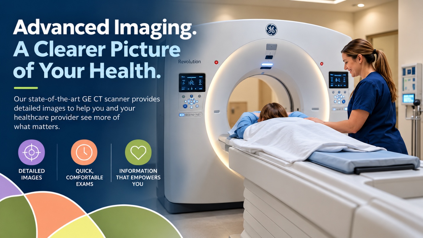
A CT scan lets us look inside the body in a way an ordinary X-ray cannot.
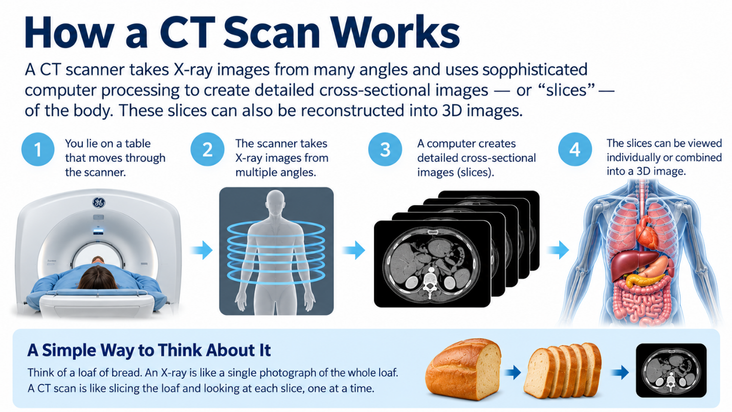
Think of a Loaf of Bread
An X-ray is incredibly useful, but generally creates a two-dimensional image in which structures can overlap.
CT—short for computed tomography—works differently.
Imagine looking at a loaf of bread from the outside.
Now imagine slicing that loaf and examining it one slice at a time.
That's roughly the concept behind CT.
The scanner collects X-ray information from multiple angles while sophisticated computer processing creates detailed cross-sectional images—or "slices"—of the body.
Those slices can also be reconstructed to help physicians examine anatomy in multiple planes and, in some applications, three dimensions.
Instead of only seeing the outside of the loaf...
we can examine what's inside.
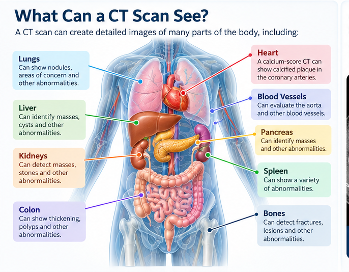
So What Can CT See?
A lot.
Depending on the type of examination, CT can create detailed images of areas including the:
Heart and blood vessels
Lungs
Liver
Kidneys
Pancreas
Spleen
Colon
Bones
Abdomen and pelvis
And other internal structures.
That's one reason CT has become such an important imaging technology throughout medicine.
But here's an important distinction:
Being able to image an organ doesn't mean CT can detect every disease affecting that organ.
No imaging technology can do that.
Different medical questions require different tests.
The Heart: Seeing Calcium in the Coronary Arteries
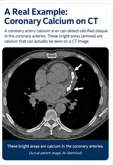
One particularly valuable application of CT is coronary artery calcium scoring.
A calcium-score CT looks for calcified plaque in the coronary arteries—the vessels that supply blood to the heart.
The result is expressed as a calcium score that can give a healthcare provider additional information about coronary artery disease and cardiovascular risk.
You can't feel a calcium score.
A stethoscope can't hear one.
And cholesterol numbers don't create a picture of your coronary arteries.
CT can provide that additional view.
The Lungs: Looking for Small Abnormalities
CT is exceptionally useful for imaging the lungs.
In fact, low-dose CT is the recommended screening test for lung cancer in people who meet established high-risk criteria.
CT creates detailed images that can reveal small lung nodules and other abnormalities that may need additional evaluation.
Most lung nodules are not cancer.
That's important.
Finding something on a scan isn't the same as receiving a diagnosis.
It means your healthcare provider has information to evaluate and determine what—if anything—should happen next.
The Abdomen: A Lot Is Happening in There
Think about how many organs are packed into your abdomen.
Liver.
Kidneys.
Pancreas.
Spleen.
Intestines.
Blood vessels.
And other structures.
CT can provide detailed cross-sectional images of these areas, helping physicians identify many kinds of abnormalities.
Depending on the exam and clinical situation, CT may be used to evaluate masses, kidney stones, inflammation, internal bleeding and other conditions.
Again, CT doesn't detect everything.
But it can provide physicians with an extraordinary amount of anatomical information.
What About Cancer?
This is where we need to be very clear.
There is no single scan that can rule out every cancer.
CT can identify many abnormalities, including some tumors, but different cancers behave differently.
Some are easier to visualize with CT than others.
Some require contrast-enhanced imaging.
Some are better evaluated with MRI, ultrasound, mammography, PET imaging, endoscopy or other technologies.
And imaging alone doesn't always determine whether an abnormality is cancer.
Sometimes additional imaging is needed.
Sometimes laboratory testing is needed.
Sometimes a biopsy is needed.
That's why we talk about imaging as information, not a guarantee.
CT and MRI Aren't Competitors
People sometimes ask:
"Which is better—CT or MRI?"
That's a little like asking:
"Which is better—a hammer or a screwdriver?"
It depends on what you're trying to do.
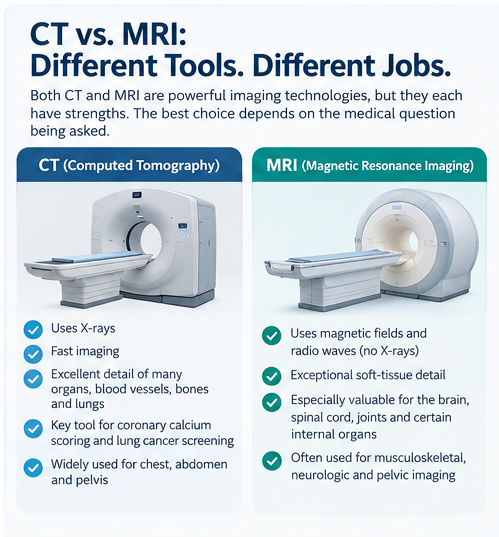
MRI provides extraordinary detail for many soft tissues and is especially valuable for certain examinations of the brain, spinal cord, joints and other areas.
CT is exceptionally fast and provides excellent detail of many organs, blood vessels, bones and lungs. It is also the established technology for coronary calcium scoring and low-dose lung cancer screening.
The goal isn't to choose the machine with the most impressive name.
It's to use the right imaging technology for the medical question being asked.
What CT Can't Do
This may be just as important as understanding what it can do.
A CT scan can't guarantee that you're healthy.
It can't detect every cancer.
It can't predict everything that will happen in the future.
It can't replace your physician.
It can't replace bloodwork.
And it doesn't replace established screening tests such as mammography or colon cancer screening when those tests are appropriate for you.
Preventive imaging is another tool.
A very powerful one.
But still a tool.
Why The Technology Matters
Modern CT scanners collect an enormous amount of imaging information very quickly.
At Virtual Imaging, we use advanced GE CT technology to create detailed images of structures inside the body.
But the machine is only part of the equation.
The examination needs to be appropriate.
The images need to be acquired correctly.
The findings need to be interpreted in the context of the examination.
And patients need to understand what happens next if something requires further evaluation.
Technology matters.
So do the people using it.
A Picture Doesn't Tell Us Everything
But sometimes it tells us something we couldn't have learned any other way.
That's the remarkable thing about medical imaging.
Bloodwork gives us numbers.
A physical examination gives us observations.
Your family and medical history give us context.
And imaging can give us something entirely different:
A picture.
Put those pieces together and you and your healthcare provider can have a much more informed conversation about your health.
Take the First Step
You don't need to become a radiologist to understand preventive imaging.
You just need to ask good questions.
What is this scan designed to look for?
Is it appropriate for someone with my age, history and risk factors?
What are its limitations?
What happens if something is found?
Those are exactly the kinds of questions informed patients should ask.
At Virtual Imaging, we believe advanced technology is most valuable when it gives people something they can actually use:
Information.
Because sometimes the difference between wondering what's happening inside your body...
and knowing more about it...
Is simply taking a look.
For more information on preventative screenings please contact Virtual Imaging at 770-730-0119 or make an appointment.
Sources and Further Reading: